


Nikon PreciPoint Scanners
The M8 and O8 slide scanners from PreciPoint are versatile all-in-one solutions that open up entirely new possibilities for your workflow. All functions are accessible via a computer touchscreen, making them fully-fledged digital microscopes. The intuitive software allows you to...
Turnkey supply, installation and training · Service across Ukraine
The M8 and O8 slide scanners from PreciPoint are versatile all-in-one solutions that open up entirely new possibilities for your workflow. All functions are accessible via a computer touchscreen, making them fully-fledged digital microscopes. The intuitive software allows you to easily navigate between specimens with just the touch of your fingers. Scan both entire slides and selected areas of interest using objectives ranging from 20x to 100x (with or without immersion oil).
PreciPoint M8—A digital microscope and slide scanner in a single device to streamline and digitize your workflow
Key Benefits
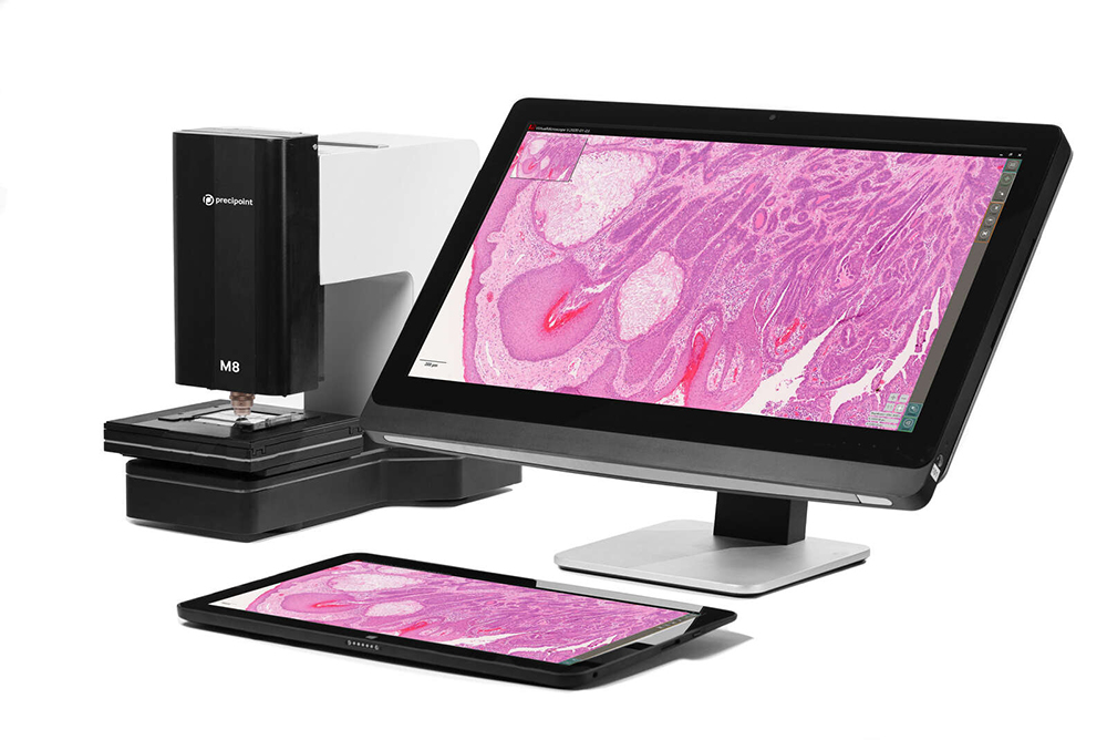
All-in-one device
The M8 and O8 integrate seamlessly into your workflow and adapt to it, accelerating your research activities.
Live Mode
Live Mode provides the capabilities of a digital microscope. Observe your sample in real time and zoom in to view different layers and structures. The overview image helps you navigate the sample.
Instant Scan Mode
Instant Scan Mode is unique to PreciPoint. It provides a large field of view for digital scanning at the speed of a microscope. Access a 15×15 mm area in high resolution in a matter of seconds. Perform analyses, identify rare events, add annotations, take notes, and digitize everything. Generate all the reports you need.
Slide Scan Mode
Get the highest-quality scans for digitizing your microscope slides. You can scan both a selected region of interest (ROI) and the entire slide (WSI), regardless of the sample’s thickness, size, or complexity (e.g., cytological or osteological slides).
Real-Time Remote Control
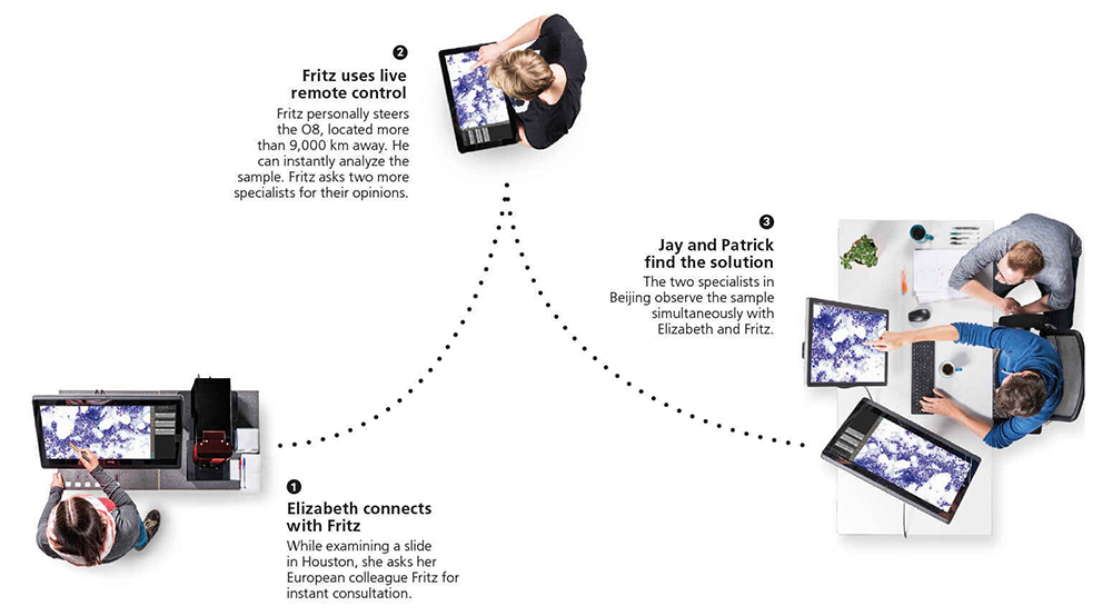
Thanks to fully automated and motorized microscopes and scanners, users can access and control the system remotely, with the option of simultaneous viewing by multiple users to obtain a second opinion. High-quality images with a wide field of view are available in seconds, and users can view different layers by manually adjusting the focal plane. Remote control is possible from computers, laptops, tablets, and even smartphones. Enhance the way you participate in training, conferences, collaboration, second-opinion consultations, and more.
Cloud Technologies and Image Analysis: PreciCloud
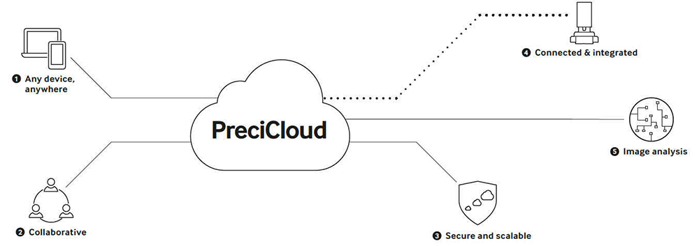
- Work seamlessly from anywhere. Store, view, share, and access digital images and specimens from anywhere in the world on any device. Our web solution works in both desktop and mobile browsers. The platform provides everything you need to organize tasks and projects of any scale.
- Set up collaborative workflows and virtual classrooms. PreciCloud’s comprehensive tools let you create virtual classrooms, organize collaborative work, and conduct consultations to get a second opinion. Experts and colleagues can collaborate simultaneously while located in different places.
- Security and scalability. PreciCloud is fully tailored to your data: users can manage access rights and permissions, as well as access logs for documentation or auditing. Our cloud platform is versatile, secure, and scalable.
- Full integration with PreciPoint products. All PreciPoint solutions are interconnected and fully integrated. You can upload scans from a PreciPoint microscope or scanner directly to PreciCloud. Share and access slides in a virtual environment without having to manually transfer files.
- Use image analysis to improve workflow efficiency. PreciPoint offers a suite of powerful automated analysis tools that utilize cutting-edge image recognition and machine learning algorithms. Preliminary analysis is performed using deep learning approaches, followed by a final review by the user.
Technical specifications
- Manufacturer
- Nikon
- Directions
- Microscopy, Histopathology
- Industries
- Educational and research laboratories, Clinical laboratory diagnostics, Medicine
- Type
- Equipment
Microscope and Scanner
| Feature | M8 | O8 |
|---|---|---|
| Operating modes adapted to various workflows | Live mode, InstantScan mode, SlideScan mode | Live mode, InstantScan mode, SlideScan mode |
| Illumination | Transmitted light; LED, bright field | Transmitted light; LED, bright field |
| Barcode scanning | Upon request | Upon request |
| Supported lenses | 20x – 60x (non-immersion) | Without immersion and with immersion oil |
| Dimensions | 45 cm × 40 cm × 30 cm; 25 kg | 45 cm × 40 cm × 30 cm; 25 kg |
| Automated microscope | Automated X-Y stage, automated Z-axis | Automated X-Y stage, automated Z-axis |
| Control | Computer-based control; real-time remote control thanks to automation | Computer-based control; real-time remote control thanks to automation |
Digitization
| Feature | M8 | O8 |
|---|---|---|
| Scanning Parameters | Full-slide imaging (WSI) or partial digitization | Full-slide imaging (WSI) or partial digitization |
| Scanning Algorithms | Various scanning algorithms adapted to specimen quality | Various scanning algorithms adapted to specimen quality |
| InstantScan mode | A large field of view in high resolution in just seconds | A large field of view in high resolution in just seconds |
| Scanning speed per specimen | 20x: 2 min | 100x: 1 hour 20x: 2 min |
| Resolution for scanning a single specimen |
60x: 0.85 NA: 0.16 μm/px 40x: 0.75 NA: 0.24 μm/px 20x: 0.50 NA: 0.479 μm/px |
100x: 1.3 NA: 0.096 μm/px 60x: 0.85 NA: 0.160 μm/px 40x: 0.75 NA: 0.240 μm/px 20x: 0.5 NA: 0.479 μm/px |
| Slide Capacity | 25 x 75 mm (2 slides) or 50 x 75 mm (1 slide) | 25 x 75 mm (2 slides) or 50 x 75 mm (1 slide) |
| Z-axis resolution | 25 nm | 25 nm |
| XY-axis resolution | 78 nm | 78 nm |
| Z-stacking | Yes | Yes |
Cloud Services, Image Analysis, and Computer
| Feature | M8 | O8 |
|---|---|---|
| Control software | MicroPoint (included) | MicroPoint (included) |
| Viewing software | ViewPoint* (included, unlimited users) | ViewPoint* (included, unlimited users) |
| Data storage | PreciCloud cloud storage for slides | PreciCloud cloud storage for slides |
| Image analysis | Several software solutions available upon request, tailored to the client's needs | Several software solutions available upon request, tailored to the client's needs |
| Computers | Various computers recommended and certified by PreciPoint | Various computers recommended and certified by PreciPoint |
| Connecting a microscope to a computer | USB 3.0 | USB 3.0 |
| Image output formats | PNG, JPEG, TIFF, BMP, VMIC, XLS, and many others | PNG, JPEG, TIFF, BMP, VMIC, XLS, and many others |
Customer reviews
There are no reviews for this product yet. Have a question about operation or configuration? A specialist will reply within 24 hours.
Purchase, delivery and warranty terms
How to order?
Click “Price request” and leave your contacts. A manager will clarify the configuration, prepare a commercial offer and answer technical questions within 24 hours.
Delivery and supply
Direct supply from the manufacturer, no intermediaries. Lead time depends on the configuration and is fixed in the offer; we deliver across Ukraine.
Warranty and service
Manufacturer's warranty on the equipment. Installation, qualification and staff training; a service engineer responds within 48 hours.
Procurement documents
Commercial offer with and without VAT, contract specification. CE, ISO, IVD certificates, declarations of conformity and user manuals.
Frequently asked questions: Nikon PreciPoint Scanners
No questions yet — ask yours via a request.